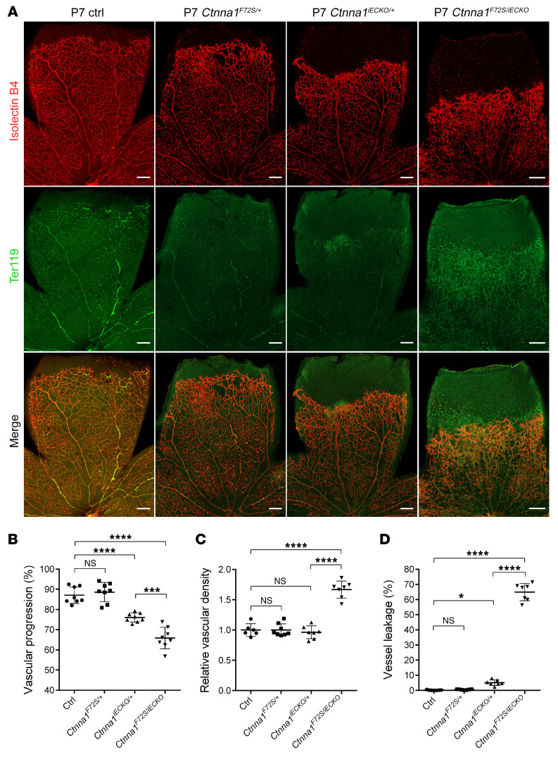
Figure 8. Ctnna1F72S/iECKO mice have retinal vasculature similar to that of Ctnna1iECKO/iECKO mice.
(A) Anti-Ter119 (green) and IB4 (red) immunofluorescence staining of retinas from P7 control, Ctnna1F72S/+, Ctnna1iECKO/+, and Ctnna1F72S/iECKO mice. Scale bars: 200 μm. (B–D) Quantification of vascular progression, relative vascular density, and vessel leakage. Error bars, SD. *P < 0.05, ***P < 0.001, and ****P < 0.0001, by 1-way ANOVA with Tukey’s multiple-comparison test (n ≥6). Experiments were performed independently at least 3 times.

