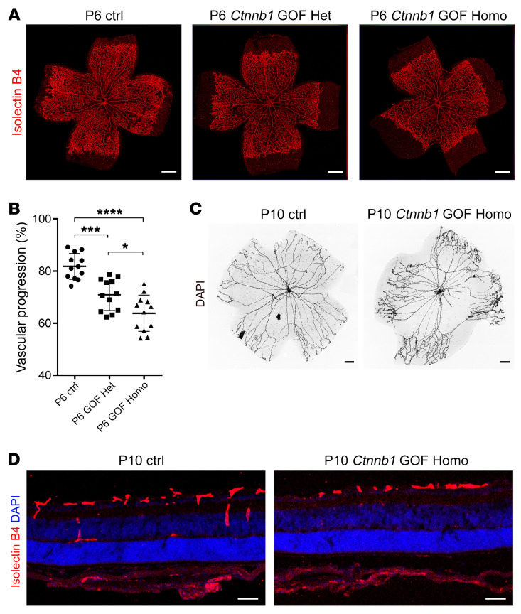
Figure 9. Ctnnb1 allele GOF in mice causes retinal vascularization defects.
(A) P6 flat-mounted retinas from control, Ctnnb1floxedexon3/+ Pdgfb-iCre-ER heterozygous (Ctnnb1 GOF Het), and Ctnnb1floxedexon3/floxedexon3 Pdgfb-iCre-ER homozygous (Ctnnb1 GOF Homo) mice were stained with IB4. Compared with those of littermate controls, retinas from the heterozygous Ctnnb1 GOF and homozygous Ctnnb1 GOF mice showed incomplete retinal vascularization. Scale bars: 500 μm. (B) Quantification of vascular progression at P6. Error bars indicate the SD. *P < 0.05, ***P < 0.001, and ****P < 0.0001, by 1-way ANOVA with Tukey’s multiple-comparison test (n = 12). (C) DAPI staining of hyaloid vessels in the eyes of control and Ctnnb1 GOF Homo mice, showing relatively delayed hyaloid vessel regression in the eyes of Ctnnb1 GOF Homo mice. Scale bars: 250 μm. (D) Frozen retinal sections from P9 control and Ctnnb1 GOF Homo mice were costained with IB4 (red) and DAPI (blue). Scale bars: 25 μm. Experiments were performed independently at least 3 times.

