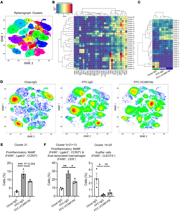
Figure 6. Anti-VCAM1Ab treatment in FFC-fed mice attenuates recruitment of proinflammatory monocytes to the liver.
CyTOF was performed on IHLs of control IgG–treated chow-fed mice and FFC-fed mice treated with either VCAM1Ab or control IgG. (A) Twenty-six unique clusters were defined by a 30 cell-surface marker panel (shown in Supplemental Table 5) using the Rphenograph clustering algorithm and were visualized on a tSNE plot. (B) Heatmap demonstrating the distribution and relative intensity of the cell-surface markers used in the clustering analysis. (C) Heatmap showing the relative abundance of each cluster for each mouse. (D) Representative tSNE plots of each experimental group. Red indicates high-frequency categorization of cells to a cluster; blue indicates low frequency. (E) Cell percentage of cluster 21 among total IHL population in mice from the different experimental groups. (F) Cell percentage of combined clusters consisting of proinflammatory MoMFs and scar-associated macrophages and those consisting of Kupffer cells. n = 3 per group. *P < 0.05; **P < 0.01; ***P < 0.001, 1-way ANOVA with Bonferroni’s multiple comparison.

