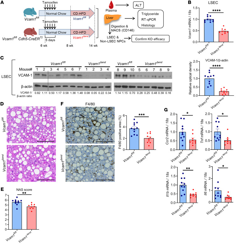
Figure 8. Loss of endothelial Vcam-1 in CD-HFD–fed mice attenuates hepatic inflammation.
Six-week-old Vcam1fl/fl;Cdh5-CreERT2 mice were intraperitoneally injected with 4 mg of tamoxifen for 5 consecutive days and used as endothelial cell–specific Vcam1 knockout (Vcam1Δend) mice. Vcam1fl/fl mice injected with the same dose of tamoxifen were used as controls. Vcam1fl/fl and Vcam1Δend mice were fed the CD-HFD starting at the age of 8 weeks for 6 weeks to induce NASH. (A) Schematic representation of the experimental model. (B) The mRNA expression levels of Vcam1 in LSECs were evaluated by real-time qPCR. FC was determined after normalization to 18S rRNA and expressed relative to Vcam1fl/fl mice. (C) Western blot for VCAM-1 protein levels in LSECs from CD-HFD–fed Vcam1fl/fl and Vcam1Δend mice. β-Actin was used as a loading control (the dotted line indicates excluded mouse due to poor protein quality) (left). Quantification of VCAM-1 protein level relative to β-actin was assessed by densitometry (right). (D) Representative images of H&E staining of liver tissues. Scale bars: 100 μm. (E) NAS. (F) Representative images of F4/80 staining of liver sections (left). Scale bars: 100 μm. F4/80-positive areas were quantified in 10 random ×10 microscopic fields and averaged for each animal (right). (G) Hepatic mRNA expression levels of Ccr2, Tnf, Il1b, and Il6 were assessed by real-time PCR. FC was determined after normalization to 18S expression and expressed relative to Vcam1fl/fl mice. n = 9 to 10 per group. Graphs represent mean ± SEM. *P < 0.05, **P < 0.01; ***P < 0.001; ****P < 0.0001, unpaired t test.

