
Fig. 1.
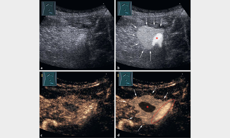
A 20-year-old male patient with acute left lower abdominal pain in the previous three days and a normal CRP value (<5 mg/l). a B-mode US image shows an echogenic, non-compressible lesion adjacent to the air-filled sigmoid colon. b Illustration of the manifestation of EA in the B-mode US image in image A. Arrowheads indicate the echogenic, non-compressible lesion as a typical B-mode US pattern of EA. The lesion is adjacent to the air-filled sigmoid colon (*). c CEUS after 33 s shows inhomogeneous enhancement with a small central area of non-enhancement, surrounded by hyperenhancement of the inflammatory fatty tissue. d Illustration of the manifestation of EA on the CEUS image in image C. Arrowheads indicate the hyperenhanced inflammatory fatty tissue. The centrally located infarcted fat tissue shows non-enhancement (*). The air-filled sigmoid colon is bordered in red.