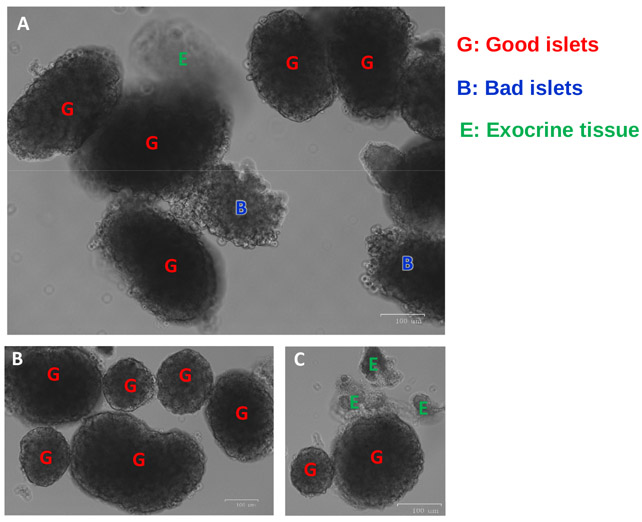
Figure 8: Representative islet images from Fluorescence Cell-Imager.
(A) Good islets, bad islets and exocrine tissue are seen. (B) A cluster of good islets is shown. (C) Panel shows undigested exocrine tissue attached to a good islet. Good/healthy islets show smooth round edges, indicated by the red letter “G”; bad/damaged islets show irregular shape and rough edges, and are indicated by blue letter “B”. Undigested exocrine tissues appear translucent, often attached to islets, and are indicated by green letter “E”.

