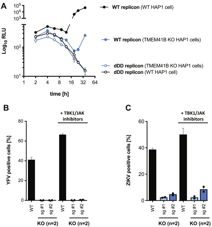
Figure S7.
TMEM41B’s Role in the Flavivirus Life Cycle, Related to Figure 7
(A) WT and polymerase dead (dDD) YFV Renilla luciferase (Rluc)-reporter replicon RNAs transfected into WT and TMEM41B KO HAP1 cells with Rluc signal plotted as relative light units (RLU) plotted across a time course of 36 h. Rluc signal increases up to 4 h as the transfected RNA is translated. Rluc signal declines between 4 and 12 h as the transfected RNA decays (dDD) or is removed from the translating pool to serve as template for viral RNA replication. Rluc signal increases in WT cells transfected with WT replicon RNA, whereas the signal is dramatically reduced in TMEM41B-deficient cells.
(B and C) WT and TMEM41B KO A549 cells infected with (B) YFV 17D, MOI = 0.025 PFU/cell for 48 h and (C) ZIKV, MOI = 0.05 PFU/cell for 48 h in the presence or absence of innate immune inhibitors. Cells were treated with 500 nM TBK1 and pan-JAK inhibitors or vehicle control (0.05% DMSO) 24 h prior to infection and throughout the experiment. The percentage of infected cells increases in WT cells in the presence of TBK1/JAK inhibitors, but not in TMEM41B KO cells. sg #1 and sg #2 indicates KO clones generated using TMEM41B sgRNA1 and sgRNA2, respectively. Two KO clones were used in each experiment as indicated in the figure. WT and KO clones were assayed in triplicate. Individual dots represent the average of the triplicates and error bars show the standard deviation of results from the average of the KO clones.

