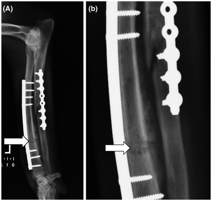
FIGURE 1.

Lateral radiograph (A) and inset (B) of a canine radius and ulna with implant‐associated osteomyelitis 1 month following repair of an open traumatic fracture. Osteolysis of the radial diaphysis (white arrow) is seen as a large, irregularly marginated, rectangular, lucent region with heterogeneous bony sclerosis that obscures the fracture margins. This abnormal region of bone is bordered caudally (between the radius and ulna) by moderate and irregularly marginated periosteal new bone formation. Similar but fainter osteolytic and osteoproliferative changes are seen surrounding the long oblique fracture within the ulna. There is also regional soft tissue swelling characterized by increased soft tissue opacity and undulating cutaneous margins. The infection resolved with prolonged antibiotic therapy based on culture and sensitivity testing and eventual removal of the plates and screws following healing of the fractures
