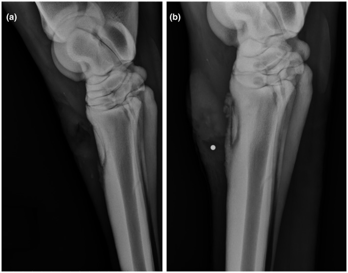
FIGURE 2.

Lateral radiographs of a Quarter Horse stallion depicting an open traumatic chip fracture of the left third metatarsal bone both 2 wk (A) and 5 wk (B) following the initial traumatic injury. After 2 weeks, only soft tissue changes associated with the original open wound and presumptive acute osteomyelitis are seen overlying the fracture dorsally. By 5 wk (B), osseous changes of chronic osteomyelitis with a sequestrum are seen, characterized by irregularly marginated periosteal new bone formation and a sharply demarcated zone of lucency and sclerosis (i.e., involucrum) surrounding the fracture fragment. A round metal opaque radiography marker was placed in a cutaneous draining tract (i.e., cloaca) overlying the sequestrum. The patient fully recovered with surgical removal of the necrotic fracture fragment, debridement, and antibiotic therapy based on culture and sensitivity testing
