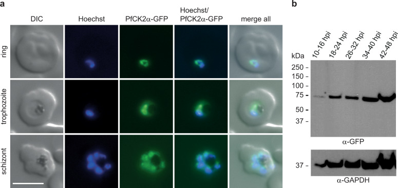
Fig. 1. Expression and localisation of PfCK2α in NF54/AP2-G-mScarlet/CK2α-GFP parasites.
a Expression and localisation of PfCK2α-GFP in ring, trophozoite and schizont stage parasites by live cell fluorescence imaging. Representative images are shown. Nuclei were stained with Hoechst. DIC, differential interference contrast. Scale bar = 5 µm. b Western blot analysis showing expression of PfCK2α-GFP at several time points during the IDC. Protein lysates derived from an equal number of parasites were loaded per lane. MW PfCK2α-GFP = 66.8 kDa, MW loading control PfGAPDH = 36.6 kDa.

