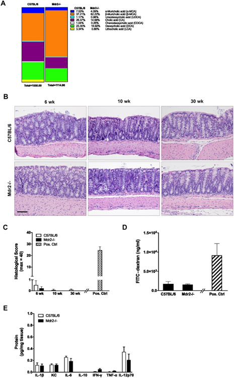
Figure 1. Baseline colonic phenotype in Mdr2−/− mice with active liver disease.
WT and Mdr2−/− mice were euthanized at age 6, 10 and 30 weeks. Feces and colon tissues were collected. (A) Levels of fecal bile acids from 10-week old WT and Mdr2−/− mice were measured using mass spectrometry. (B, C) Representative colon histology (hematoxylin and eosin [H&E]; 200X) and histological scores. Positive control in panel C is WT/DSS from Fig. 2E. (D) Colonic barrier function in 10-week old mice was assessed by FITC-dextran gavage. Serum levels of FITC-dextran were measured four hours post gavage. Positive control is WT/DSS from Fig. 2G. (E) Colon tissue was homogenized and cytokine protein levels were measured. n ≥ 4 mice per group. Data are expressed as means ± SEM, two-way analysis of variance.

