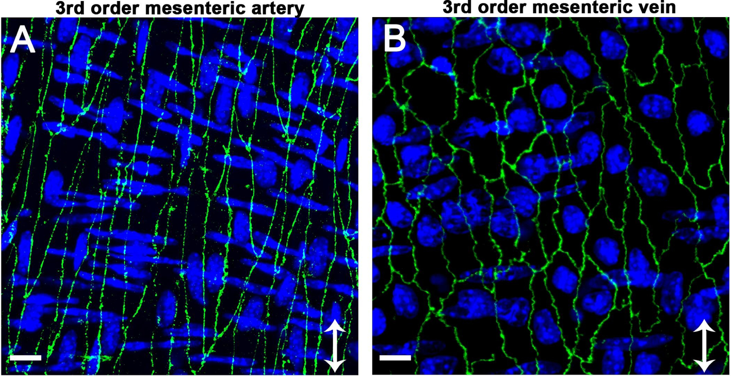
Figure 4. Polarization of endothelium in response to different flow environments.

Claudin-5 (green, Thermofisher 34–1600) is used to demonstrate differences in polarization of interendothelial proteins to lateral edges of the cells in a murine third order mesenteric artery with high flow (A) compared with a murine third order mesenteric vein with low flow (B). The images also highlight endothelial morphological differences in arteries versus veins: arterial endothelial cells are elongated in the direction of flow while venous endothelial cells are more rectangular in shape and resemble cultured cells. For each image, arrow indicates direction of flow, scale bar is 10μm, and nuclei are shown in blue, with endothelial nuclei oriented vertically and SMC nuclei oriented horizontally. Images were obtained with a Zeiss 880 LSM Airyscan module (40x oil objective) and are shown as Z-projections.
