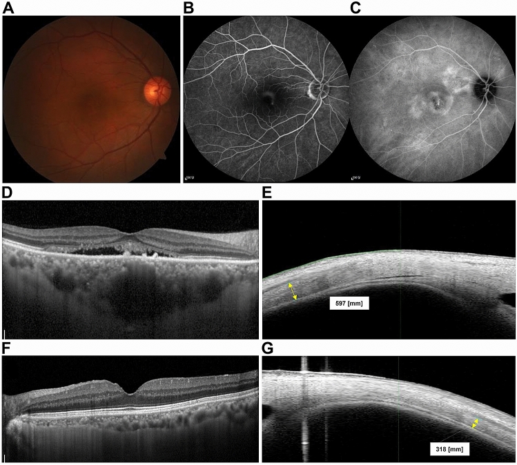
Figure 3.
Representative cases of CSC eyes (A–E) and normal eyes (F,G). Color fundus photographs of CSC eyes show serous macular detachment (A) and fluorescein angiography shows typical smoke stack pattern of leakage (B). Late phase image of indocyanine angiography shows multiple areas of choroidal hyperpermeability. We observed very thick choroid in enhanced-depth imaging (EDI) of optical coherence tomography (OCT) and relatively thick sclera in swept source anterior segment (AS) OCT in CSC eyes (D,E), compared with those of normal eyes (F,G).

