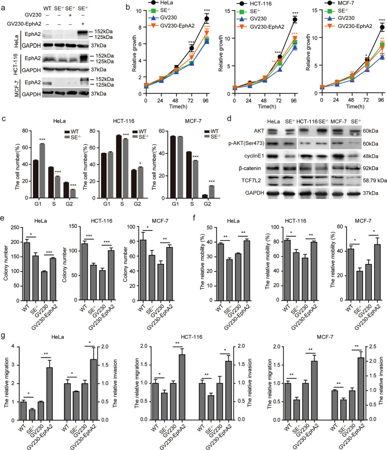
Fig. 4. EphA2-SE knockout inhibited proliferation and migration of HeLa, HCT-116, and MCF-7 cells.
a Expression level of EphA2 as measured by western blotting in Hela, HCT-116 and MCF-7 cells. The overexpression vector GV230-EphA2 was constructed to express EphA2-EGFP fusion protein, and then transfected into the EphA2-SE homozygous deletion SE−/− cell lines. b Cell proliferation was measured using MTT assay after EphA2-SE knockout and EphA2 overexpression. c Flow cytometry was used to detect the cell cycle of HeLa, HCT-116 and MCF-7 cells. d Effect of EphA2-SE deletion on PI3K-AKT signaling pathway. e The summary graphs of the number of colonies counted. f The migration distance was calculated. g Migration and invasion efficiency measured by transwell assay. Error bars, mean ± SD. n ≥ 3. p values were calculated using t-test. *p < 0.05, **p < 0.01, ***p < 0.001. WT: wild type cell; SE−/−: EphA2-SE homozygous deletion; GV230 SE−/−: Cells transfected with empty vector GV230; GV230-EphA2-SE−/−: Cell lines overexpress EphA2.

