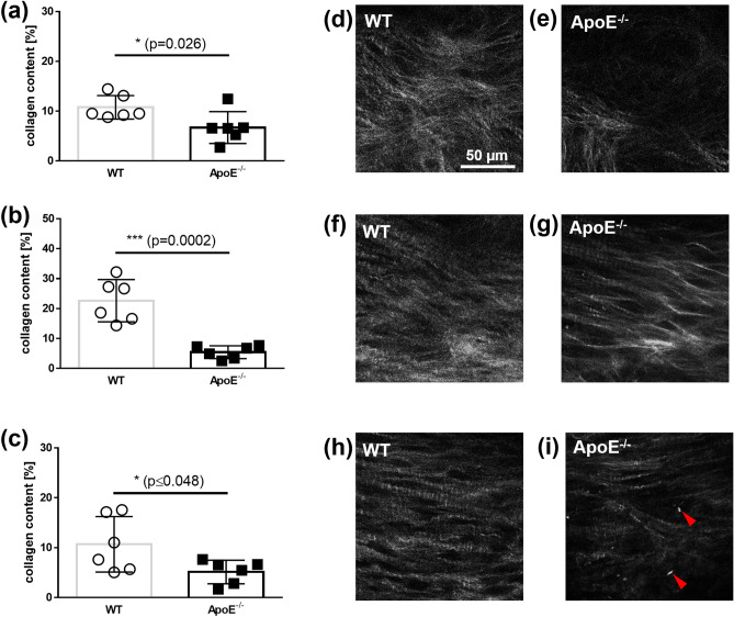
Figure 3.
Quantitative analysis of the collagen content in a leaflet using SHG images. The mean collagen content from WT mice and ApoE−/− mice from (a) the region 1; from (b) region 2; and from (c) the region 3. Representative SHG images of the region 1 of the aortic valve leaflets from (d) WT mice and (e) ApoE−/− mice. Representative SHG images of the region 2 from (f) WT mice and (g) ApoE−/− mice. Representative SHG images of the region 3 of (h) WT mice and (i) ApoE−/− mice, acquired in the middle of the thickness. The dimensions of all images are 140 µm × 140 µm.

