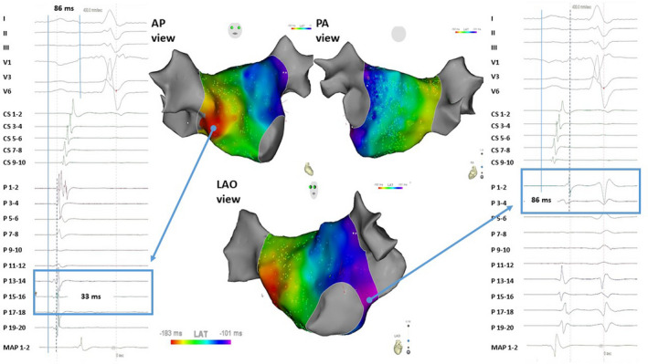
Figure 1. Electroanatomical mapping of a left atrial in a patient with a short P wave (86 ms) in anterio‐posterior, postero‐anterior, and left anterior oblique view.

The earliest left atrial breakthrough occurred in the mid septum 33 ms after P wave onset as determined on surface ECG; the local electrogram and activation is visible on the catheter mapping (Pentaray – P 15‐16) of the right panel. Latest left atrial activation is on the lateral side of the left atrium close to the mitral valve ring; the local electrogram and activation is visible on the catheter mapping (Pentaray – P 1‐12) of the left panel. AP indicates antero posterior view; LAO, left anterior oblique view; MAP 1‐2, distal electrode pair of the ablation catheter; P, Pentaray mapping; PA, postero anterior view.
