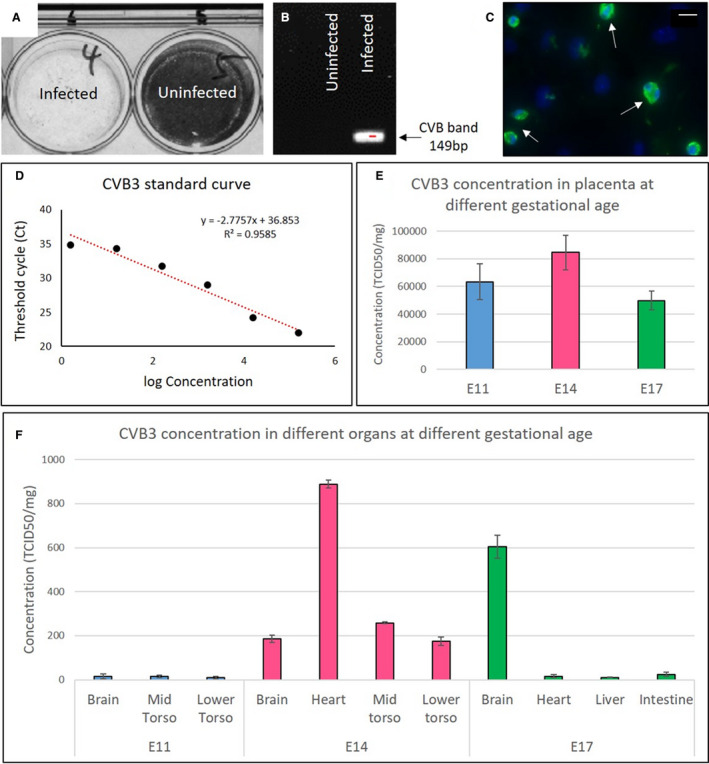
Figure 1. Vertical transmission of coxsackievirus B (CVB) 3 from infected pregnant dams to the fetus.

A, The presence of infectious virus in fetal lysates from infected and uninfected dams (embryonic day [E] 9) was analyzed using a cell culture infectivity assay; the lack of stained cells indicates the presence of virus. B, Reverse transcription–polymerase chain reaction (PCR) with enterovirus (EV)–specific primers, showing amplification of the 149‐bp (determined by agarose gel electrophoresis) virus band only in the tissue lysate of a fetus from an infected dam (at E9). C, Immunohistochemistry using anti‐EV antibody on cultured fetal heart (~E14) explanted at E11 from a dam infected at E9 (white arrows). Virus quantification by quantitative PCR. D, Standard curve based on the concentration of CVB3 stock. E, Virus present in placenta at E11, E14, and E17 after infection at E9 (n=6 for each gestational age). F, Virus present in fetal organs at E11, E14, and E17 after infection at E9 (n=6 for each gestational age). To quantify the amount of virus in tissue samples, the standard curve equation was used along with its coefficient of determination (R 2). Bar=20 µm. TCID50 indicates 50% tissue culture infective dose.
