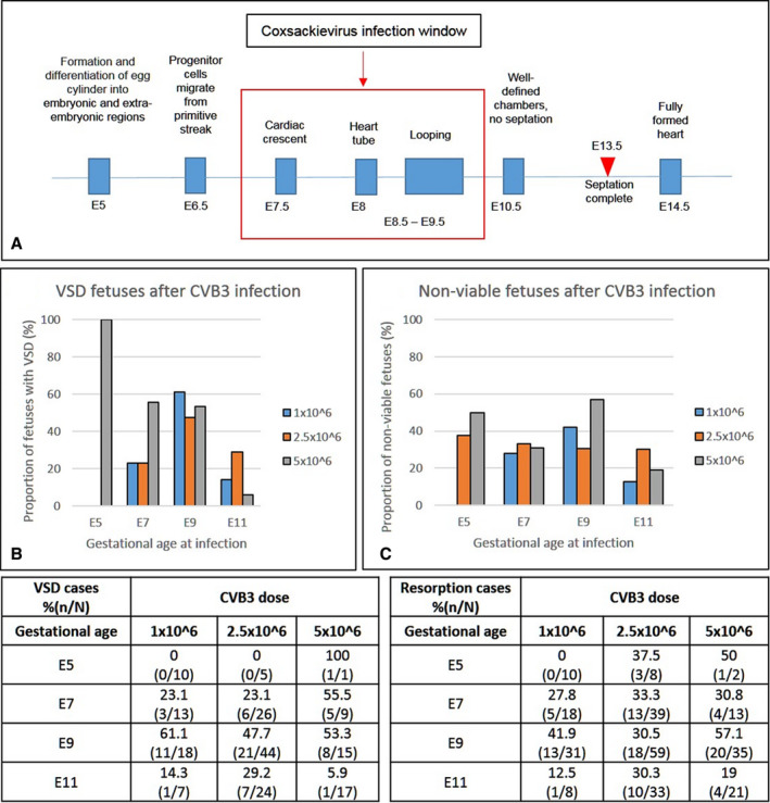
Figure 3. The effect of viral dose and gestational age at infection on the incidence of ventricular septal defect (VSD) and fetal demise.

A, Schematic representation of the timeline of heart development in the mouse and critical window for coxsackievirus B (CVB) 3 infection. B and C, Dams were infected at various stages of gestation (embryonic day [E] 5, E7, E9, or E11) with different doses of virus (1.0, 2.5, and 5.0 × 10⁶ 50% tissue culture infective dose). Graphs and their corresponding tables show the percentage of fetuses with VSDs (B) and nonviable (dead or resorbed) fetuses (C) along with the total examined in each experimental group. The χ2 test was used to analyze statistical significance between control and test groups by calculating P values.
