
Figure 7. Mouse model of coxsackievirus B (CVB) 3 infection during pregnancy and proposed mechanism for ventricular septal defect (VSD) formation after infection.
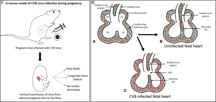
I, Mouse model of CVB virus infection during pregnancy, leading to vertical transmission of virus from infected pregnant mice to the fetus, resulting in 3 outcomes: fetal death, congenital heart defect, and no cardiac phenotype. IIa–c, The proposed mechanism of VSD generation. Normal intraventricular septum (IVS) formation (a), with the proliferation of the muscular IVS and the outflow tract septum, resulting in a fully formed septum (b), is shown (proliferating cardiomyocytes are shown in green). c, In CVB‐infected fetuses, cell proliferation is suppressed, which leads to incomplete septum formation and VSD. LA indicates left atrium; LV, left ventricle; RA, right atrium; and RV, right ventricle.