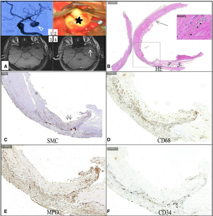
Figure 4.

A man had a middle artery aneurysm (triangle arrow).
(A1) with a uniform atherosclerosis appearance (star) on intraoperative imaging (A2), which had significant enhancement (arrow) on postcontrast vessel wall magnetic resonance imaging (A4). A hematoxylin and eosin stain (B) showed the thickened aneurysm wall contained hemosiderosis (star) and calcification (triangle). A vascular smooth muscle cells (SMC) stain (C) indicated hyperplasia and derangement of SMCs within the aneurysm wall (double arrow). Clustered inflammatory cells, including CD68+ macrophages (D), myeloperoxidase+ (MPO+) leukocytes (E), and T and B lymphocytes, were noticed within the aneurysm wall, and CD34 stain (F) indicated plenty of neovascularization within the aneurysm wall.
