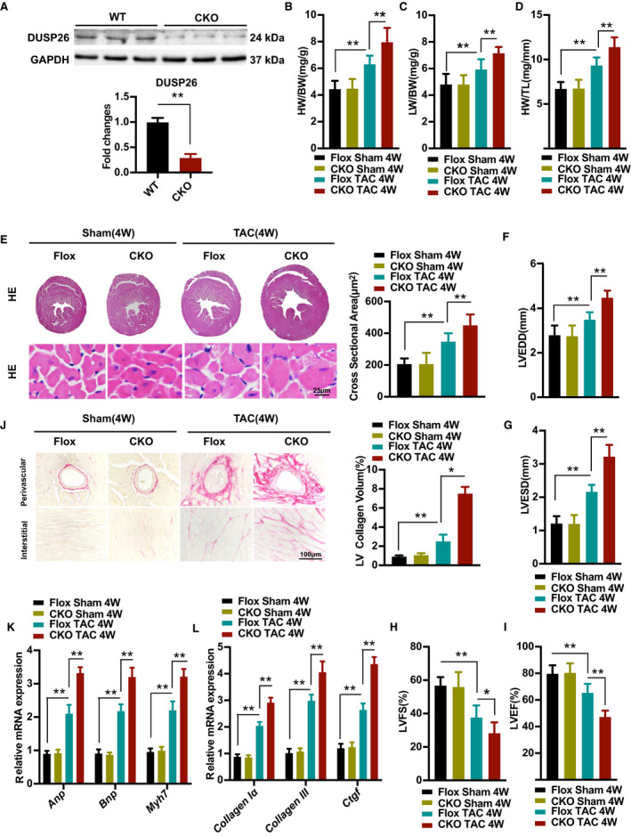
Figure 3. Cardiac‐specific DUSP26 deletion aggravated pressure overload–induced hypertrophy in vivo.

A, Western blot analysis of protein expression of DUSP26 in hearts of 8 week‐old WT or DUSP26 knockout mice (n=3 per group). B through D, The ratios of heart weight to body weight (HW/BW; B), lung weight to body weight (LW/BW; C) and heart weight to tibia length (HW/TL; D) in Flox and DUSP26‐CKO mice after 4 weeks of TAC or sham surgery. (n=10 per group). E, Representative images of heart sections with histological examination and quantification of myocyte cross‐sectional area in Flox and DUSP26‐CKO mice after 4 weeks of TAC or sham surgery as indicated by groups (scale bar, 25 μm; n=6 per group). F through I, Echocardiographic measurements of indicated groups. Average data of left ventricular end‐diastolic diameter (LVEDD; F), left ventricular end‐systolic diameter (LVESD; G), left ventricular fractional shortening (LVFS; H) and left ventricular ejection fraction (LVEF; I) (n=10 per group). J, Representative images of histological analysis of cardiac perivascular and interstitial fibrosis and quantification for the fibrotic area in different genotype groups. (scale bar, 100 μm; n=6 per group). K and L, Real‐time PCR analysis of mRNA levels of multiple hypertrophic marker genes (Anp, Bnp, and Myh7) (K) and fibrotic marker genes (collagen Iα, collagen III, and Ctgf) (L) in indicated groups (n=4 per group). Data are presented as the mean ±SD. *P<0.05, **P<0.01. Anp indicates atrial natriuretic peptide; Bnp, brain natriuretic peptide; BW, body weight; CKO, conditional knockout; Ctgf, connective tissue growth factor; DUSP26, dual‐specificity phosphatase 26; Myh7, myosin heavy chain 7; and TAC, transverse aortic constriction.
