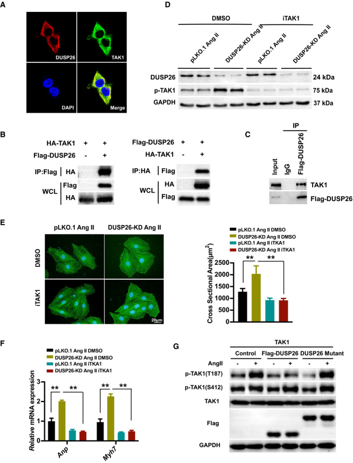
Figure 6. DUSP26 bound to TAK1 and blocked its phosphorylation to attenuate DUSP26‐regulated cardiac hypertrophy.

A, Representative images of colocalization of DUSP26 and TAK1 proteins in HEK293T cells with double immunofluorescent staining. (blue, nucleus; green, TAK1; red, DUSP26). B, Total protein extracted from HEK293T cells cotransfected with HA‐TAK1 and Flag‐DUSP26 to perform immunoprecipitation for examining the interaction between TAK1 and DUSP26. C, Total protein extracted from H9C2 cells infected with Flag‐DUSP26 to perform coimmunoprecipitation experiment for examining the interaction between DUSP26 and endogenous TAK1. D, Western blot analysis of protein levels of p‐TAK1 and DUSP26 in H9C2 cells infected with viruses targeting DUSP26 or nontargeting pLKO.1 vector and then treated with iTAK1 (2.5 μmol/L) or DMSO along with angiotensin II (1 μmol/L) stimulation for 48 hours. E, Representative images of H9C2 cells infected with lentiviruses targeting DUSP26 or nontargeting pLKO.1 vector and then treated with iTAK1 (2.5 μmol/L) or DMSO and angiotensin II (1 μmol/L) stimulation for 48 hours. Cardiomyocytes were stained with antibody against α‐actinin. (blue, nucleus; green, α‐actinin; scale bar, 20 μm). F, Quantification results of mRNA levels of the hypertrophic marker genes (Anp, Myh7) in cardiomyocytes infected with viruses targeting DUSP26 or nontargeting pLKO.1 vector and then treated with iTAK1 (2.5 μmol/L) or DMSO and angiotensin II (1 μmol/L) stimulation for 48 hours. G, Western blot results of the phosphorylation protein levels of TAK1 at T187 and S412 and total protein levels of TAK1 in H9C2 cells infected with Flag‐DUSP26 or DUSP26 mutant and treated with angiotensin II (1 μmol/L) or PBS for 30 minutes. **P<0.01. Anp indicates atrial natriuretic peptide; ERK, extracellular signal‐regulated kinase; iTAK1, TAK1‐specific inhibitor; JNK, c‐Jun N‐terminal kinase; Myh7, myosin heavy chain 7; p38, p38 kinase; and TAK1, transforming growth factor‐β activated kinase 1.
