
Figure 1. Echocardiographic images of a 79‐year‐old symptomatic female with a long‐standing history of hypertension and a picture consistent with hypertrophic obstructive cardiomyopathy.
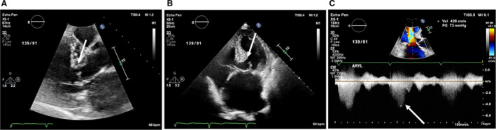
A, Parasternal long‐axis image demonstrating a sigmoid‐shaped upper septal bulge with concomitant systolic anterior motion of mitral valve. (B) Four‐chamber image demonstrating a sigmoid‐shaped upper septal bulge with concomitant systolic anterior motion of mitral valve. (C) Continuous Doppler across the left ventricular outflow tract demonstrating severe dynamic obstruction.