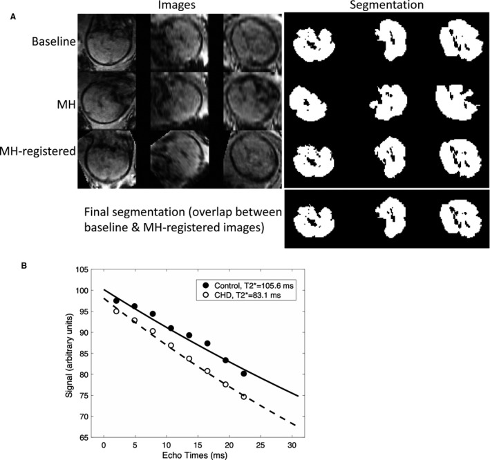
Figure 1. T2* fetal brain MRI.

A, Images from 3 orthogonal plans were segmented and registered to obtain the final segmentation for T2* measurements at baseline and with MH; B, T2* decay curves for a control and CHD subject with hypoplastic left heart syndrome. The gestational age at fetal MRI was 33 3/7 weeks in the control subject and 34 2/7 weeks in the CHD subject. CHD indicates congenital heart disease; MH, maternal hyperoxia; and MRI, magnetic resonance imaging.
