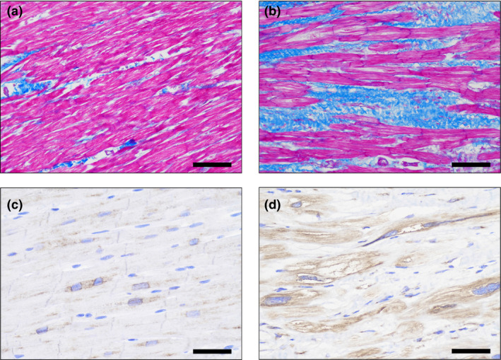
Figure 1. Representative histologic staining from septal biopsies in patients with HOCM.

The left panel corresponded to representative pictures of Masson's trichrome staining (A) and immumohistochemical staining (C) for LOX in a control subject, respectively. The right panel corresponded to representative pictures of Masson's trichrome staining (B) and immumohistochemical staining (D) for LOX in a HOCM patient, respectively. Myocardial fibrosis was stained in blue (A, B), and the LOX expression was stained in brown (C, D). Magnification ×200. HOCM indicates obstructive hypertrophic cardiomyopathy; and LOX, lysyl oxidase.
