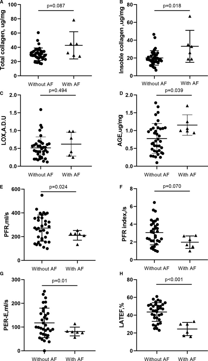
Figure 3. Comparisons between patients with HOCM with AF and those without AF for molecular and cardiovascular magnetic resonance indices.

Compared with patients without AF, patients with AF showed higher myocardial total collagen (A), insoluble collagen (B), and AGE expression (D), but insignificant LOX expression (C). In addition, patients with AF also exhibited lower PFR (E), PFR index (F), PER‐E (G), and LATEF (H). ADU indicates arbitrary densitometric units; AF, atrial fibrillation; AGE, advanced glycation end‐product; HOCM, hypertrophic obstructive cardiomyopathy; LATEF, total left atrial ejection fraction; LOX, lysyl oxidase; PER‐E, early peak emptying rate; and PFR, peak filling rate.
