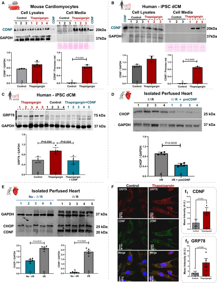
Figure 1. Thapsigargin (TG) treatment increases the levels of CDNF in human and mouse cardiomyocytes as well as its secretion to the extracellular media.

A, Mouse cardiomyocyte or (B) Human iPSC‐dCM were treated for 20 hours with TG (1 µmol/L) and the CDNF contents in cell lysates and cell media were evaluated by Western blot analyses. C, Levels of GRP78 in cell extracts of hiPSC‐dCM cultures treated with TG (1 µmol/L) or with CDNF (1 µmol/L) before TG. D, Levels of CHOP from hearts subjected to I/R or I/R after CDNF treatment (1 µmol/L/5 minutes). E, Levels of CHOP and CDNF from rat hearts subjected to I/R protocol in relation to no‐I/R condition. F, Levels of CDNF and GRP78 in mouse cardiomyocytes before and after TG treatment, measured by confocal imaging. Scale bar represents 20 µm (f1 and f2) immunocytochemistry quantification of CDNF and GRP78, respectively. The levels of CDNF, GRP78, and CHOP were normalized to GAPDH levels in cell lysate and to Ponceau red in extracellular media. Recombinant CDNF was used as control. Each lane represents a different cell culture. The protein expression values are expressed as mean±SEM with 1‐way ANOVA followed by Bonferroni post hoc test. CDNF indicates cerebral dopamine neurotrophic factor; CHOP, CCAAT/‐enhancer‐binding protein homologous protein; iPSC‐dCM, iPSC‐dCM, induced pluripotent stem cells differentiated into cardiomyocytes; GRP78, 78 kDa glucose‐regulated protein; and I/R, ischemia/reperfusion.
