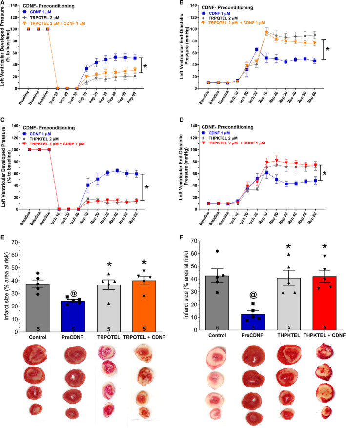
Figure 6. The cardioprotective effect of exoCDNF is blocked by heptapeptides that bind to KDEL‐R.

Time courses of left ventricular developed pressure (LVDP) (A and C) and left ventricular end‐diastolic pressure (LVEDP) (B and D) during I/R protocol. CDNF (1 µmol/L), peptide (in [A, B, and E] TRPQTEL and in [C, D and F], THPKTEL L; 2 µmol/L of each peptide) or CDNF (1 µmol/L)+peptide (2 µmol/L) were perfused before ischemia (5 minutes). E and F, Infarct area of hearts. Representative cross‐section images of TCC‐stained ventricle hearts subjected to I/R in the absence or in the presence of CDNF and peptides. Number in each column is n of hearts. The data were expressed as means±SEM. @ P<0.01 vs control; *P<0.01 vs preCDNF. With 1‐way ANOVA followed by Bonferroni post hoc tests for infarct area analysis and 2‐way ANOVA followed by Bonferroni post hoc tests for LVDP and LVEDP analysis. CDNF indicates cerebral dopamine neurotrophic factor; isch, ischemia; rep, reperfusion; and TCC, triphenyltetrazolium chloride.
