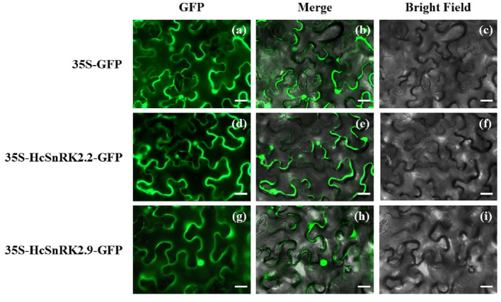
Figure 8. Subcellular localization of HcSnRK2.2 and HcSnRK2.9 proteins.
HcSnRK2.2-GFP and HcSnRK2.9-GFP fusion vectors were transformed into N. benthamiana leaves and subcellular localization was carried out 48 h after infiltration using Leica DM RXA2 upright fluorescent microscope. The bar indicates 20 µm.

