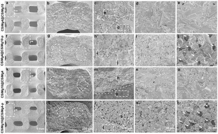
Figure 2.
SEM Images of side-wall fracture surface of the core-shell-strut scaffolds of CSiMg4@CSiMg10 (a–e), CSiMg4@CSiMg10-p (f–j), CSiMg10@CSiMg4 (k–o) and CSiMg10@CSiMg4-p (p–t). the dotted curves represent the interface of the core/shell component and the arrows indicate the formation of microporous structures in the shell layer.

