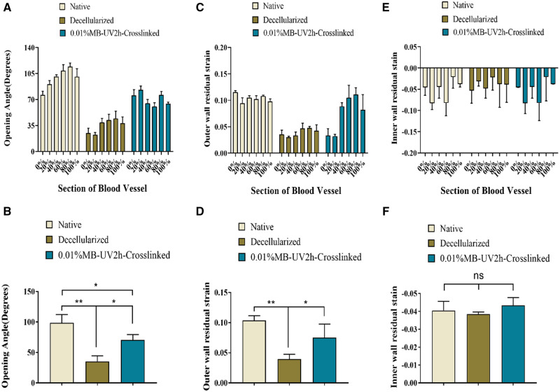
Figure 5.
Comparison of biomechanical parameters among native, decellularized and 0.01% MB-UV2h-crosslinked arteries. (A) Opening angles of crosslinked arteries appeared to increase compared with decellularized arteries. (C) The outer wall residual strains increased though crosslinking. (E) The residual strain on the inner wall did not appear to change significantly. (B), (D) and (F) reveal whether opening angles, outer wall residual strains and inner wall residual strains have a significant difference among native, decellularized and 0.01% MB-UV2h-crosslinked arteries.*Corresponds to a P < 0.05; **Corresponds to a P < 0.01; ns, not significant

