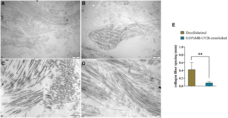
Figure 7.
TEM of decellularized and 0.01%MB-UV2h-crosslinked arteries. Example images are shown for (A) decellularized and (B) 0.01%MB-UV2h-crosslinked arteries. Zoomed-in images of (C) decellularized and (E) 0.01%MB-UV2h-crosslinked arteries showed details of collagen fibers. The comparison of spacing of collagen fibers between decellularized and 0.01%MB-UV2h-crosslinked arteries (E)

