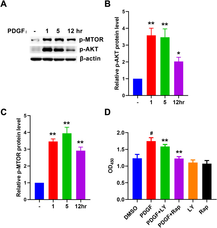
Figure 6.
PDGF induced ASM cells proliferation by activating AKT/mTOR pathway. (A–C) ASM cells were treated with PDGF (10ng/mL) for 1h, 5h, and 12h. The expression levels of p-AKT and p-mTOR were analyzed by Western blotting. The relative protein levels of P-AKT and P-mTOR were calculated by Image J, respectively and expressed as mean ± SD (n=3) of each group. * P < 0.05 and ** P < 0.01 vs control group. (D) ASM cells were treated with PDGF (10ng/mL) and LY294002 (10 μM), Rapamycin (500 nM) for 5h, respectively. Cell proliferation was detected by CCK-8. The data were expressed as mean ± SD (n=8) of each group, ** P<0.01 vs PDGF group, # P<0.01vs control group.

