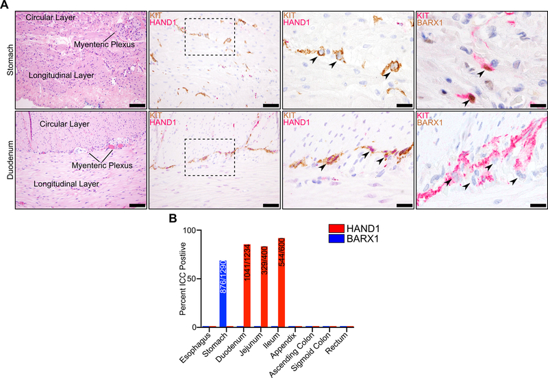
Figure 6. Anatomic and spatial restriction of HAND1 and BARX1 expression in ICC.
A, H&E staining of normal tissue with muscular layers and myenteric plexus indicated (left, scale bar 100 μm) in stomach (top row) and duodenum (bottom row). KIT (brown) and HAND1 (red) IHC at the myenteric plexus (second from left, scale bar 50 μm) with individual ICC indicated by an arrowhead (third from left, scale bar 20 μm). KIT (red) and BARX1 (brown) IHC at the myenteric plexus (right, scale bar 20 μm). B, Percentage of ICC expressing positive for HAND1 or BARX1 in normal gastrointestinal tissues. Inset values indicate the numerator of positively expressing ICC over the denominator of total ICC. For quantification, 100 ICC were evaluated per section, where possible, in at least 7 sections per anatomic location.

