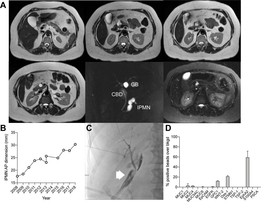
Figure 5. Enlarging IPMN concerning for high grade dysplasia in a 57 year old patient.
(A) A predominantly branch duct IPMN was monitored annually with MRI over a decade. (B) During this time, the size of the IPMN increased. (C) The patient underwent a pancreaticoduodenectomy. The postoperative course was prolonged by a bile leak which was treated by percutaneous biliary drainage. The pathology results showed a low grade IPMN. (D) Note that the MUC5AC EV levels at the time of surgery were low indicative of a low grade lesion, suggesting that surgery and complicated postoperative course could have been avoided by EV analysis.

