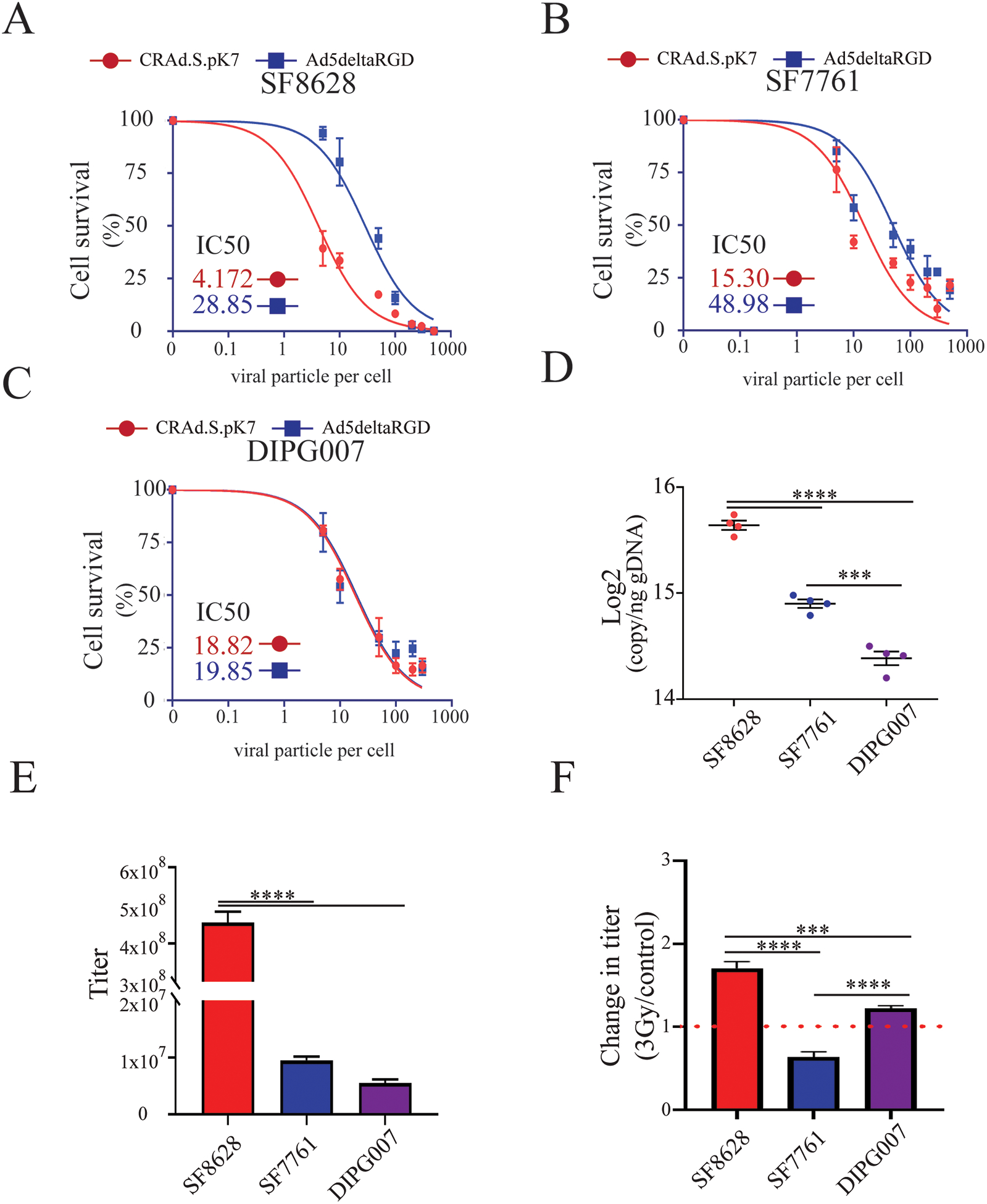
Figure 3. Oncolytic activity of CRAd.S.pK7 in patient-derived DIPG cells.

A) Kinetics of killing by CRAd.S.pK7 and Ad5deltaRGD of SF8628 DIPG cells expressing firefly luciferase were measured by the loss of luciferase activity 3 days after treatment of cells with OV (n=4). B) Kinetics of killing by CRAd.S.pK7 and Ad5deltaRGD of SF7761 DIPG cells (n=4) and C) DIPG007 DIPG cells (n=4) were assessed as described in A. D) Binding assay of CRAd.S.pK7 with the surface of DIPG cell lines SF8628, SF7761, DIPG007 was performed to assess the difference in the binding of viral particles to the cell surface using PCR-based quantification of viral DNA (One-way ANOVA with Tukey’s post hoc test, n=4. Levels of significance are: *** p <0.001 and **** p<0.0001). E) The release of CRAd.S.pK7 progeny from SF8628, SF7761, and DIPG007 DIPG cell lines was assessed by determining viral titer using staining for hexon viral protein in 293T cells (Negative binomial model was fitted and followed by Comparisons of Group Least Squares Means with Tukey-Kramer adjustment, n=4). F) Change in the progeny release of CRAd.S.pK7 from in DIPG cells after 3Gy irradiation treatment (One-way ANOVA with Tukey’s post hoc test, n=4. Levels of significance are: * p <0.05, ** p <0.01, *** p <0.001 and **** p<0.0001).
