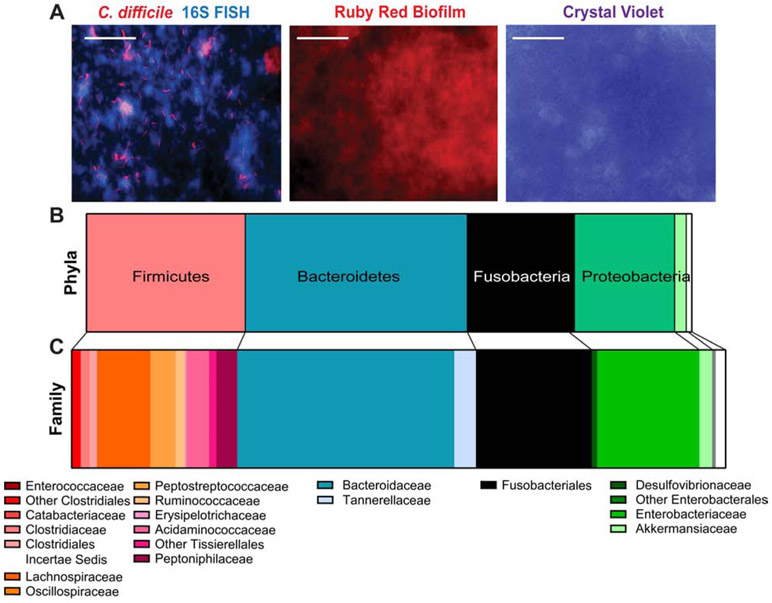
Figure 2: C. difficile interacts with other mucus-associated microbes in bioreactor mucin-based biofilms.
Bioreactors were modified with a hanging insert containing MUC2-APTS-coated cover slips. Bioreactors were seeded with stool from healthy volunteers (2x donors, 3x fecal pools, n =4) and treated with clindamycin (250 μg/mL) followed by C. difficile (106 CFU). MUC2 coated inserts were examined for biofilm production, C. difficile localization and 16S rRNA sequencing. A. FISH immunostaining for C. difficile (red) and total 16S rRNA genes (all bacteria; blue). Biofilms on coverslips were stained with ruby red biofilm tracer and crystal violet staining (scale bar = 50 μm) (representative image; n=4/stain). The composition of mucin-coated inserts was examined by 16S sequencing which revealed a unique microbial community at the phylum (B) and family (C) level.

