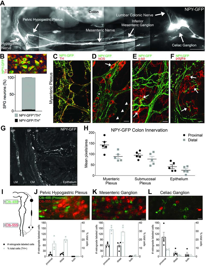
Figure 1. Individual sympathetic postganglionic neurons (SPNs) innervate proximal or distal colon.
(A) Montage shows innervation from sympathetic prevertebral ganglia to colon in NPY-GFP reporter mouse. (B) Quantification of sympathetic ganglion neurons that express NPY-GFP and/or TH (all NPY-GFP+ cells expressed TH). (C) Whole-mount immunolabeling reveals colocalization of TH and NPY-GFP+ fibers. (D) NPY-GFP+ fibers appear to innervate NOS+ (arrow) and NOS- myenteric neurons (asterisk); NPY-GFP+ varicosities are also closely opposed to NOS+ terminals within smooth muscle (arrowheads). (E-F) NPY-GFP+ fibers make close contacts to c-kit+ interstitial cells of Cajal (E) and PDGFRA+ cells (F) in the myenteric plexus. (G) Cross section of NPY-GFP mouse colon shows innervation to all layers; LM, longitudinal muscle, MP, myenteric plexus, CM, circular muscle, SP, submucosal plexus, M, mucosa, E, epithelium. (H) Comparison of NPY-GFP fiber density in proximal and distal colon. (I) Colon back-labeling experimental design. (J-L) Individual sympathetic postganglionic neurons in the pelvic hypogastric plexus (J), inferior mesenteric ganglion (K), and celiac ganglion (L) project to proximal (green) or distal (red) colon regions. Bar graphs show total number (left axis, black) and percentage (right axis, gray) of labeled cells. Scale bars, 1mm (A), 100μm (G), 20μm (C-F,J-L).

