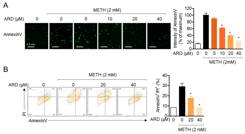
Figure 7.
(A) SH-SY5y cells (5 × 10³/well, 96-well plate) stained with 1 × AnnexinV staining reagent were pre-treated with the indicated concentration (0 to 40 μM) of aromadendrin for 1 h and exposed to 2mM METH for 24 h. AnnexinV was detected by IncuCyte® imaging system, and the intensity of AnnexinV was calculated. (B) SH-SY5y cells were pre-treated with the indicated concentration (0 to 40 μM) of aromadendrin for 1 h and exposed to 2 mM METH for 24 h. After collection, AnnexinV/PI apoptosis assay was performed by flow cytometry, and AnnexinV/PI-positive cells were presented in a bar graph. White bar indicates 0.2 mm. The mean value of three experiments ± SEM is presented. * p < 0.05 between METH-treated cells.

