
Figure 5. Removal of miR-31 suppression of Wnt1 results in shorter DVC length while Wnt1 overexpression results in extra skeletal rudiments and exogastrulation.
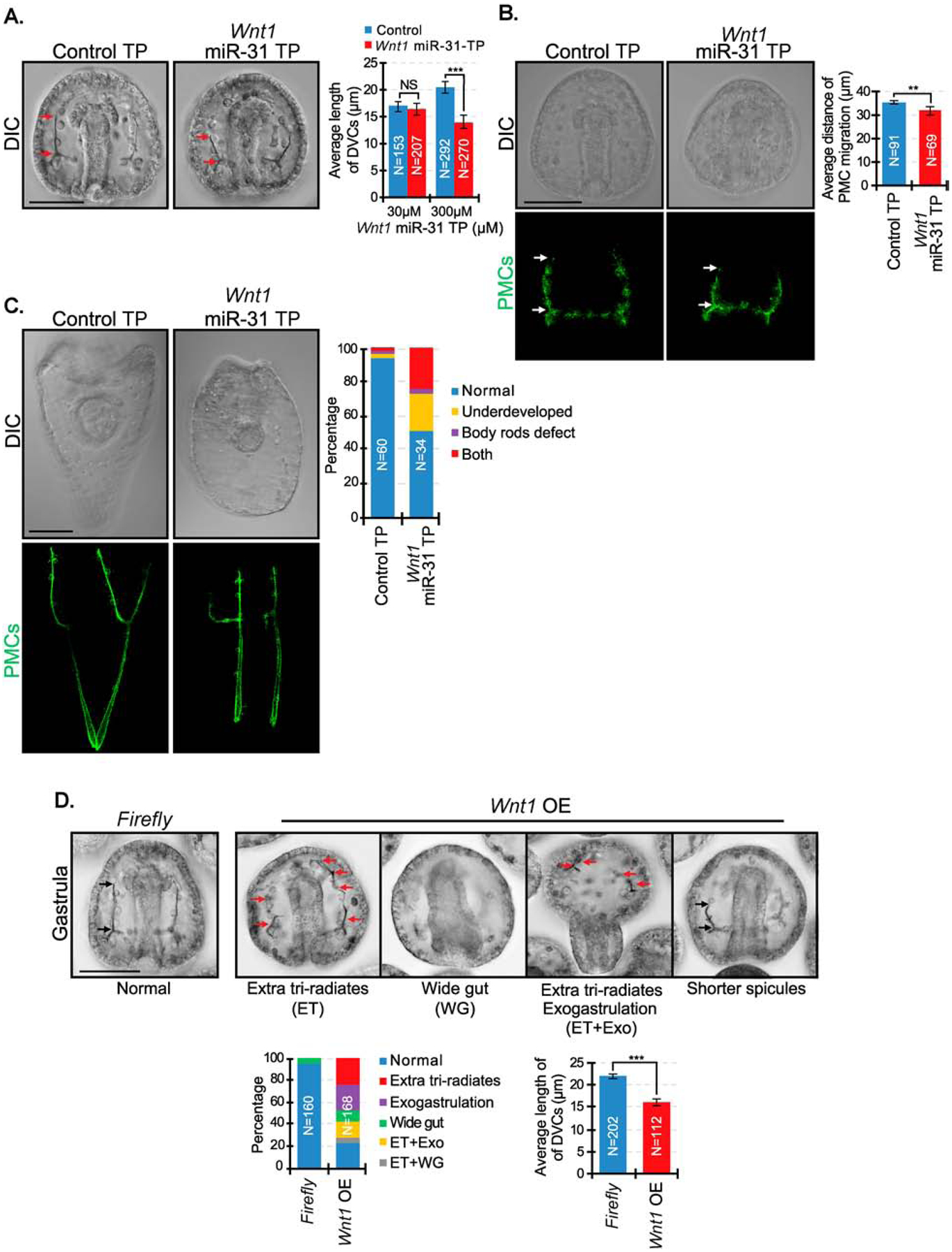
(A) Wnt1 miR-31 TP injected gastrulae (48 hpf) had decreased DVC length in a dose-dependent manner. Red arrows indicate the length of DVCs. P-value was analyzed using Student’s t-test. 2–3 biological replicates. NS=not significant. N is the total number of spicules examined. (B) Embryos were immunolabeled with PMC antibody, 1D5. PMC anterior migration is decreased in Wnt1 miR-31 TP injected embryos compared to the control injected embryos. P-value was analyzed using Student’s t-test. 2 biological replicates. White arrows indicate PMC migration distance. (C) Wnt1 miR-31 TP injected larvae (5dpf) appeared rounder with body rods that failed to meet at the posterior end compared to the control TP. 2 biological replicates. Maximum intensity projection of Z-stack confocal images are presented for the PMC patterning. (D) Overexpression of Wnt1 CDS resulted in multiple developmental defects. Red arrows indicate skeletal tri-radiates. Black arrows indicate the length of DVCs. P-value was analyzed using Student’s t-test. 4 biological replicates. N is the total number of spicules examined. Tri-radiates were counted through a series of Z-stack images. **p<0.001, ***p<0.0001. All error bars represent SEM. Scale bar = 50μm. N is the total number of embryos examined except where otherwise stated.