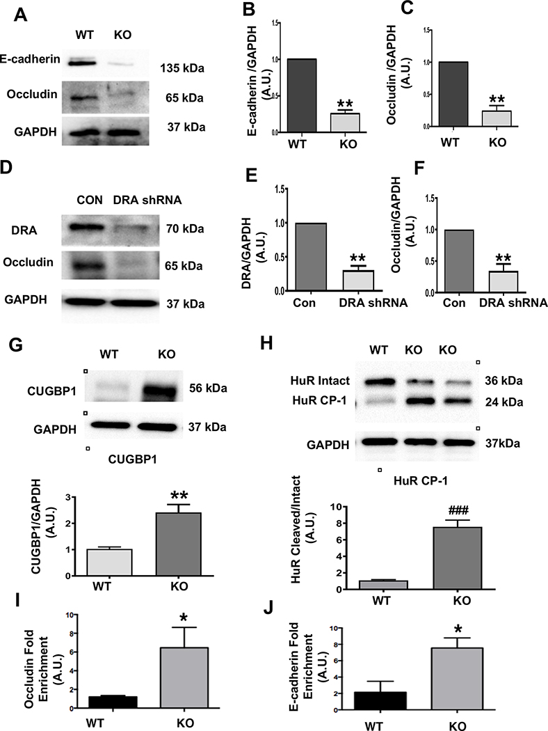
Figure 4: Loss of DRA results in reduced TJ/AJ protein expression in colonoids and Caco-2 cells and increased CUGBP1 expression, HuR protein cleavage, and binding of CUBGP1 to 3’ UTR of occludin and E-cadherin in DRA KO mice:
Protein expression of E-cadherin and occludin in colonoids from wild type and DRA KO mice by western blotting (Figure A). Densitometric analyses of band intensities (GAPDH was used as the internal control) are shown (Figures B and C). DRA and occludin immunoblotting in lentiviral shRNA mediated knockdown of DRA in Caco-2 cells are shown (Figure D). Densitometric analyses of band intensities (GAPDH was used as the internal control) are shown (Figures E and F). Values (for figures 4B,4C,4E,4F) are means ± SEM from blots from 3–4 independent experiments. Data was analyzed by unpaired t-test (**p <0.01 vs. wild type). Western blot and densitometric analysis of CUGBP1 and HuR protein levels in distal colonic mucosal lysates of wild type or DRA KO mice (Figures G and H). Results represent means ± SEM of 3–4 mice (**p <0.01 compared to wild type, ###p <0.001, cleaved compared with intact HuR). CUGBP1 binding (over IgG) to 3’UTR of occludin and E-cadherin as measured by RNP-IP Assay (Figures I and J). Values are means ± SEM of blots of 4–5 independent experiments. Data was analyzed by unpaired t-test (*p <0.05 compared to wild type).

