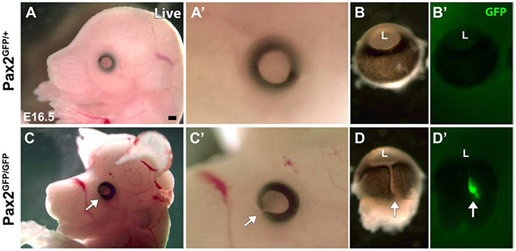
Figure 2. Brain and eye deformities of Pax2GFP/GFP embryos.
(A,A’,C,C’) Lateral views of E16.5 live embryos. Pax2GFP/GFP animals with exencephaly and ocular colobomas (arrow in C,C’). (B,B’,D,D’) The optic fissure remains open as a ventral cleft in Pax2GFP/GFP mutant eyes (arrow in D), with GFP fluorescence visible (arrow in D’) (n=4/genotype). L – lens, Rostral is left in A,C; distal up in B,D. Scalebar = 500 μm

