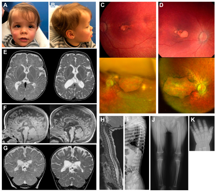
Figure 1.
Clinical images of the patient. Frontal (A) and lateral (B) photographs of the patient’s head show facial appearances with mild coarsening and a deep nasal bridge. Retinographies of the left (C) and right (D) eyes present macular colobomatous involvement due to a retinal degeneration. Upper images were obtained at the age of six months whereas lower images were taken at age two years. Cerebral magnetic resonance imaging (MRI) (E–G) show hypomyelinating leukoencephalopathy, and progressive brain and cerebellum atrophy. Left images were obtained at the age of six months, whereas right images were obtained at age two years. Lateral spine MRI image and radiograph (H,I, respectively) present small epiphyseal changes due to a spondylo-epiphyseal dysplasia at six months of age. Skeletal radiographs (J,K) show severe ossification delay when the patient was 20 months old.

