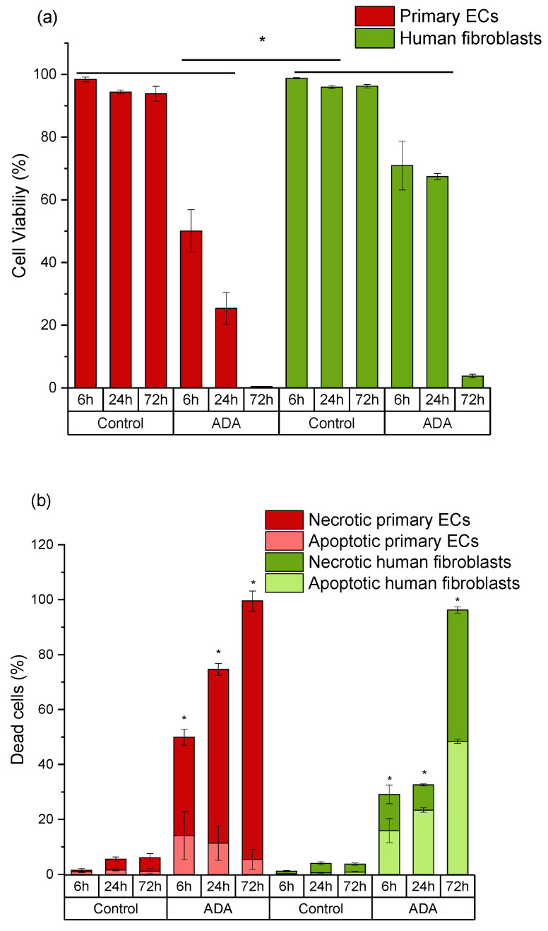
Figure 4.
Time-dependent comparison of primary endothelial cells (ECs) and fibroblasts grown in ADA. (a) Cell viability (DiI-positive cells); (b) apoptotic populations in total death cluster (DiI-negative, PI-negative staining) and necrotic populations in total death cluster (DiI-negative, PI-positive staining). Control: cells with DCFH-DA grown on plastic. * p < 0.05 indicates significant differences between the groups in cell viability (a) and number of apoptotic cells (b).

