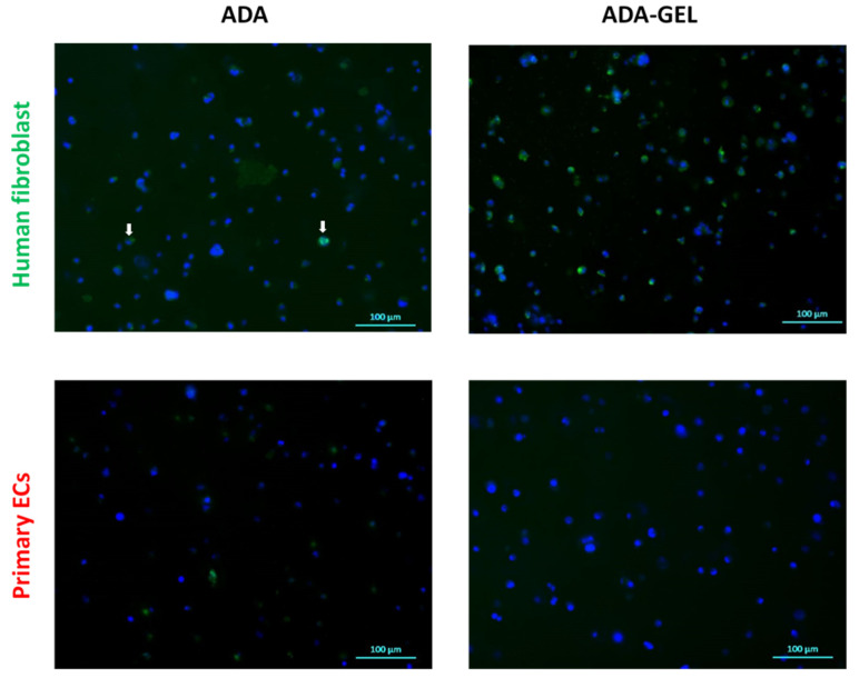
Figure 8.
Immunofluorescence staining of proliferating cells (green channel) after 72 h of incubation. Representative images show Ki-67 expression in primary ECs and human fibroblast grown in ADA and ADA-GEL. Nuclei: Hoechst (Blue), Ki-67: Alexa Fluor 488 (green). The white arrows indicate scarce fibroblasts in ADA positive for Ki-67.

