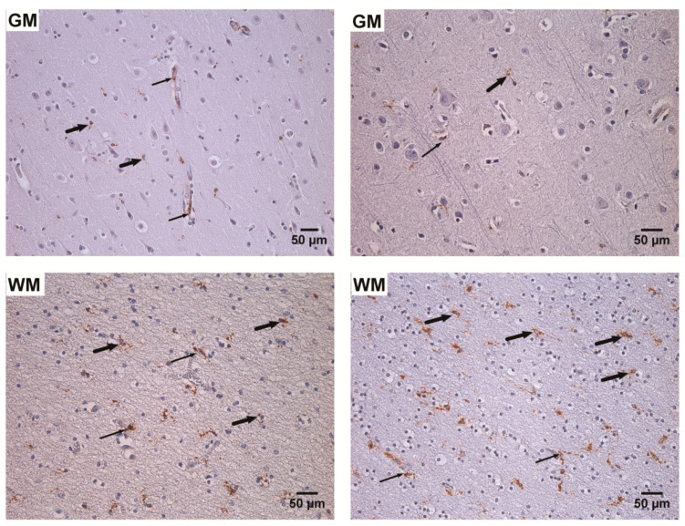
Figure 8.
Detection of CD68 positive cells by routine immunohistochemistry (IHC), CD68 immunopositive products—brown. Left: frontal gray (GM) and white (WM) matter of the unspecified encephalopathy (UEP) subject demonstrating brown reaction products in the activated microglia/macrophages (perivascular (thin arrows) and diffuse (thick arrows) location), (200×, 250×); right: temporal gray (GM) and white (WM) matter of the UEP subject demonstrating brown reaction products in the activated microglia/macrophages (perivascular (thin arrows) and diffuse (thick arrows) location), (250×, 250×).

