Figure 4.
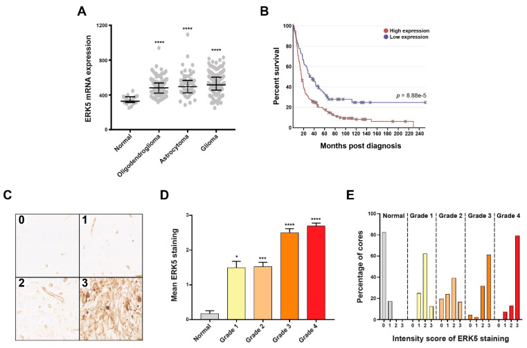
Primary brain tumours exhibit heightened ERK5 expression which is associated with tumour grade and poor patient survival. (A): Scatter plot showing relative ERK5 mRNA expression in the indicated tissue samples derived from the REMBRANDT online brain tumour repository of 524 samples. (B): Kaplan–Meier survival curves for brain tumour patients within the REMBRANDT repository differentiated for either low or high relative ERK5 mRNA expression along with the associated Chi-squared significance. (C): Representative bright field images of immunohistochemical staining showing the indicated graded intensity of ERK5 protein expression/staining of FFPE brain tumour tissue sections from the Sheffield Royal Hallamshire Hospital cohort. (D): Average intensity scoring of ERK5 protein staining within the indicated tumour grade across the 190 samples scored, which consisted of 28 normal brain, 16 grade 1, 64 grade 2, 31 grade 3, and 51 grade 4 gliomas. (E): Breakdown of the percentage of FFPE cores exhibiting the indicated staining intensity scores within normal brain samples and each tumour grade for the patient cohorts shown in panel D. Statistical significance was calculated using the Kruskal–Wallis H-test: * = p < 0.05, *** = p < 0.001, and **** = p < 0.0001 comparing the indicated tumour grade cohort to the normal brain cohort.

