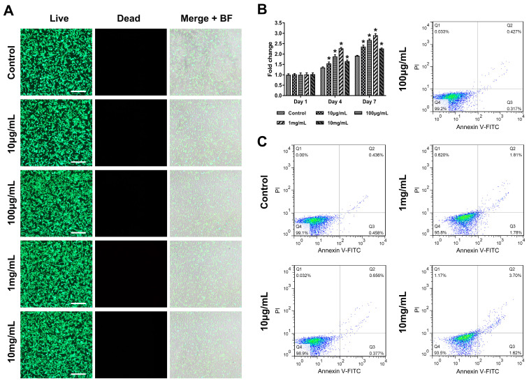
Figure 3.
The biocompatibility of rGO@Ge. (A) The live/dead staining of ADSCs cultured with different rGO@Ge concentration media, Merge+BF: the merge of Live, Dead and Background Field images, Bar: 500 μm. (B) CCK8 assay on the ADSCs proliferation at 1, 4 and 7 days. * p < 0.05 indicates a significant difference compared to the corresponding control group at each time point. (C) Flow cytometry analysis of the ADSCs apoptosis with the different rGO@Ge concentration stimulus.

