Figure 5.
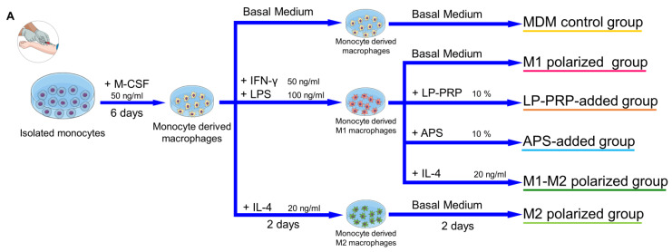
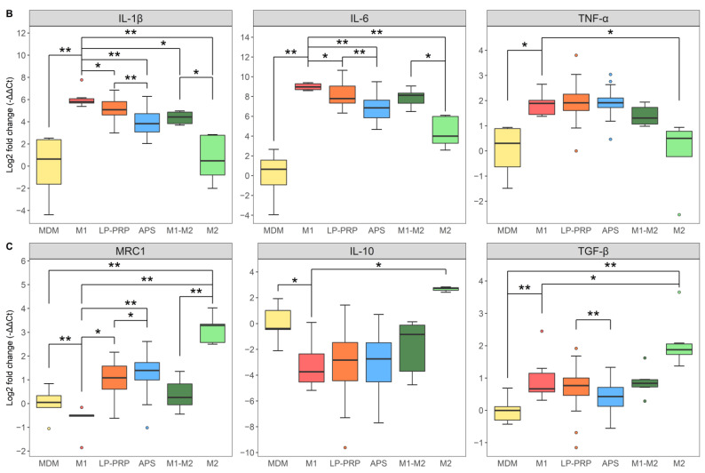
Effect of PRPs on M1 macrophages. (A) After CD14 + monocytes were isolated by the same method as described in Figure 3, they were cultured in a basal medium supplemented with 10% FBS containing M-CSF at 37 °C under 5% CO2. After six days, the media was replaced by fresh basal medium supplemented with 10% FBS containing IFN-γ + LPS, and the cells were cultured for another two days to polarize them to M1 macrophages. The medium was removed, the basal medium supplemented with 10% FBS containing supernatants obtained from LP-PRP or APS was added, and the cells were cultured for another two days. (B) Expression of M1 macrophage markers (IL-1β, IL-6, TNF-α). (C) Expression of M2 macrophage markers (MRC1, IL-10, TGF-β). Data were analyzed through qRT-PCR. -ΔΔCt values were calculated using GAPDH as an internal control. MDM control and M1 polarized groups served as negative controls; M1-M2 and M2 polarized groups served as positive controls. MDM control, M1 polarized, M1-M2 polarized, M2 polarized groups: 6 monocyte donors, 1 experiment, n = 6 per group; LP-PRP- and APS-added groups: 6 monocyte donors, 12 PRP donors, n = 72 per group. * p <0.05, ** p <0.01.


