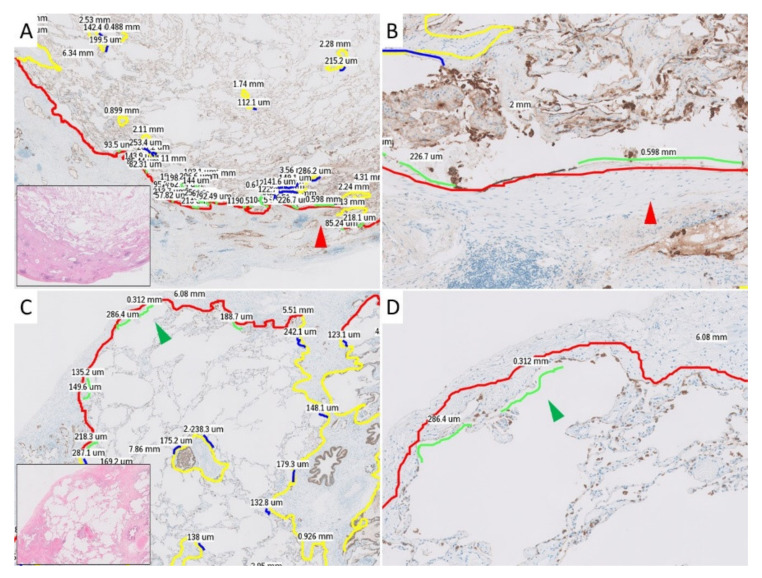
Figure 1.
Histopathological images with annotations used in the current study. The red and green arrowheads each indicate the same location. The details of the colors in the annotation lines are as follows: red, subpleural zone; yellow, paraseptal zone; green, alveolar epithelial denudation (AED) in the subpleural zone; blue, AED in the paraseptal zone. (A): Annotated pleuroparenchymal fibroelastosis (PPFE) case in middle power view. (B): Annotated PPFE case in high power view. (C): Annotated idiopathic pulmonary fibrosis (IPF) case in middle power view. (D): Annotated IPF case in high power view.

